Advanced Cone Beam Imaging
We Provide Cone Beam CT Scans in Bend, OR
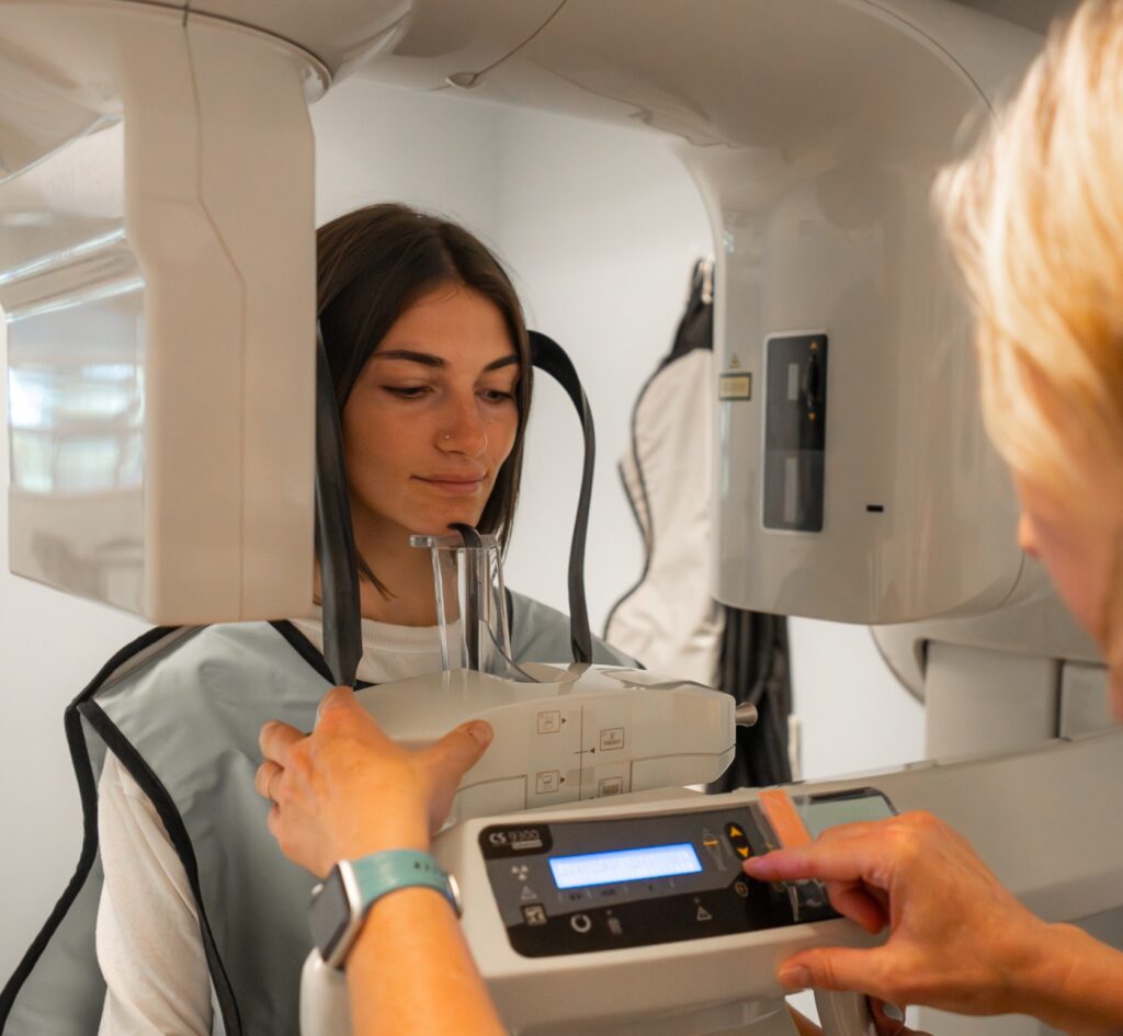
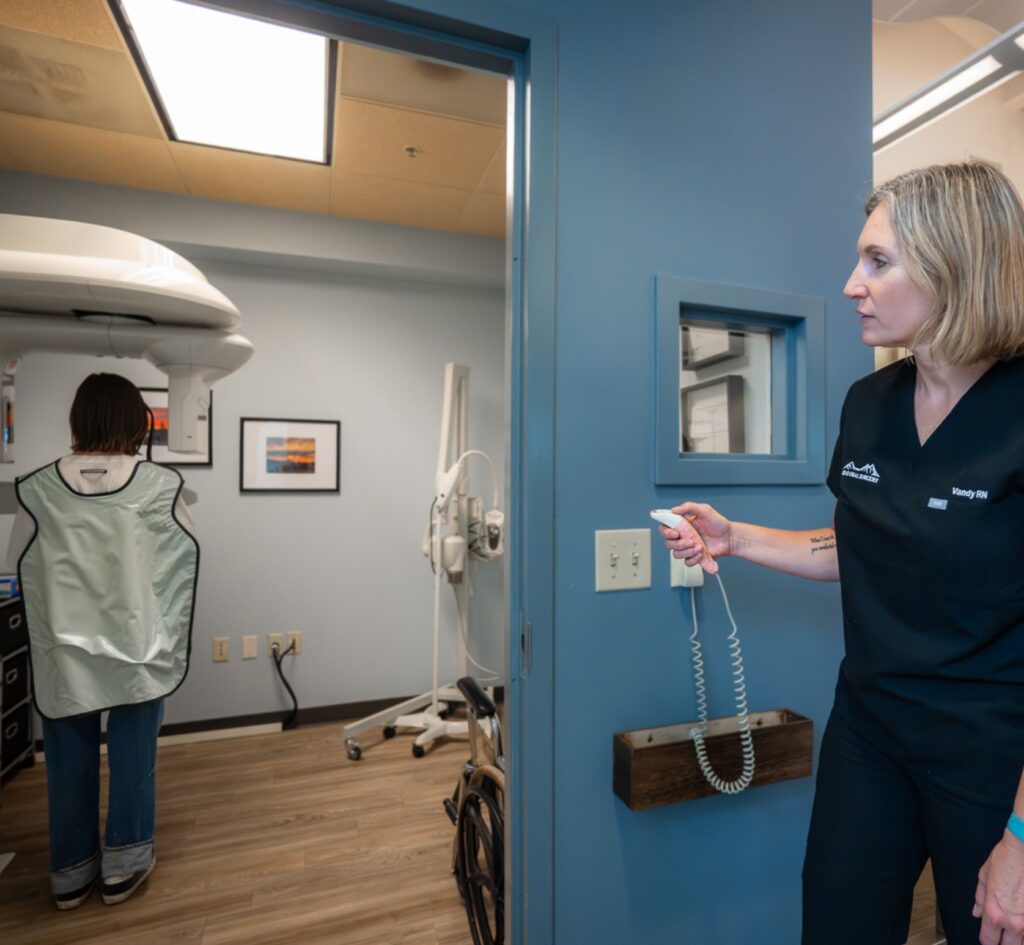
Bend Oral Surgery’s Kodak 9000/9500 Cone Beam CT Scanner evaluates and analyzes critical anatomy for implants and oral surgery within minutes – right in our office.
It gives precise, cross-sectional slices of any desired location in the upper or lower jaws, which provides the surgeon with exact anatomical information, including dimensions and locations for your particular case. This technology ensures that we have the most information available regarding your personal surgical needs.
How Does Kodak 9000/9500 Cone Beam CT Scanning Work?

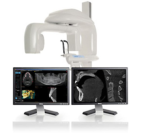
The Kodak 9000/9500 Cone Beam Computed Tomography (CBCT) system captures high-resolution 3D images of the teeth, jaw, and surrounding structures, providing detailed diagnostics and precision treatment planning for oral and maxillofacial procedures.
Step-by-Step Process:
- Patient Positioning – The patient stands or sits in the scanner, and the machine is adjusted to ensure proper alignment.
- Rotational X-ray Capture – The CBCT scanner rotates 360 degrees around the patient’s head, capturing multiple images from different angles.
- 3D Image Reconstruction – Advanced software compiles the images into a detailed 3D model, allowing surgeons to examine teeth, bone, nerves, and soft tissue in high definition.
- Diagnosis & Treatment Planning – The 3D scans help in evaluating bone density, detecting pathology, planning dental implants, assessing wisdom teeth impactions, and screening for TMJ disorders.
Advanced Diagnostics and Precision Treatment Planning
We use advanced cone beam imaging to help determine tooth positions, visualize impactions, and assess their proximity to nerves, sinuses, and bone structures.
Patients with Temporomandibular Disorder (TMD) also benefit from detailed imaging that allows for precise condyle measurements and identification of abnormalities.
Additionally, it aids in early detection of pathological issues, including bone deformities, cysts, tumors, and jaw disease, ensuring timely and effective treatment.
FAQ - Cone Beam Imaging
Cone Beam CT imaging is a specialized 3D scanning technology that captures high-resolution images of your teeth, jaw, nerves, sinuses, and surrounding facial anatomy in a single rotation. At Bend Oral Surgery, we use the Kodak 9000/9500 CBCT system, which rotates 360 degrees around the patient and compiles the captured data into a detailed 3D model. This gives Dr. DeLisi exact anatomical measurements and cross-sectional slices of any location in the upper or lower jaws, right in our office. Learn more: https://bendoralsurgery.com/advanced-cone-beam-imaging/
Our advanced cone beam imaging supports a wide range of procedures, including dental implant planning, wisdom teeth removal, impaction assessment, TMJ screening, and detection of pathological conditions such as bone deformities, cysts, and tumors. For implant cases, it provides precise bone density and nerve position data. For TMD patients, it allows detailed condyle measurements that support accurate diagnosis and treatment planning.
Accordion Conte Yes. CBCT technology focuses imaging specifically on the head and neck region rather than the full body, which significantly reduces unnecessary radiation exposure compared to traditional medical CT scans. The system delivers substantially more clinical detail than standard dental radiographs while keeping exposure minimal. The imaging is widely used in oral and maxillofacial surgery for early detection of pathology, trauma assessment, and surgical navigation. Learn more about our advanced diagnostics and screening.